EYE IMAGE
STRUCTURE OF EYE:
IT’S WORKING AND PROBLEMS
The eyeball is a spherical structure having 2.5 diameters. Structurally, the eyeball has three layers:- sclerotic layer (outer layer); choroid (middle layer) and retina (inner layer).
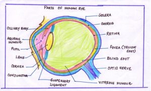
PARTS OF THE HUMAN EYE
- Outermost layer contains sclera and cornea
- Middle layer contains choroid, ciliary body and iris.
- Innermost layer consists of retina
AN OVERVIEW OF STRUCTURE, LOCATION, FUNCTIONS OF VARIOUS PARTS OF THE EYE
| PARTS OF THE EYE | STRUCTURE | LOCATION | FUNTION
|
| Sclera | Opaque fibro-elastic capsule; transparent in front at cornea | Outermost covering part of eye | i)Maintains shape of eyeball
ii)Protects inner eye |
| Lacrimal gland | Saclike structure | Upper eyelid towards the outer corner of eye. | i)Secretions wash away dust particles
ii)Lubricates eye iii)lysozyme present in tear kills the germs |
| Cornea | Transparent buging part | Front part of eye | i)Allows the light to pass into the eye.
ii)Helps in focusing. |
| Conjunctiva | Thin transparent layer | Present over the cornea. | Protects cornea. |
| Choroid | Pigmented layer containing numerous blood vessels. | Present in middle layer beneath the sclera. | i)Prevents reflection within eye.
ii)Provides eye. |
| Ciliary body | Contains ciliary muscle and blood vessels. | Present at the junction of sclera and the cornea. | Helps to maintain shape of eye. |
| Ciliary muscle | Contains a complex set of smooth muscles in a circular form. | Present over suspensory ligaments | The contraction of ciliary muscles results in spherical shape of the lense and the relaxation in the flattened shape. |
| Suspensory ligaments | Thread-like ligaments | Attached to the ciliary body to the lens. | They hold lend in position. |
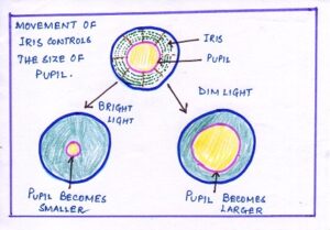
| Iris | Pigmented circle of muscular diaphragm. | Attached to the ciliary body. | It controls the size of pupil for entry of amount of light. |
| Pupil | Small aperture or opening of eye. | Present in the centre of pupil. | It helps in entering light into the eye. |
| Retina | Stretched light sensitive screen-like structure containing two types of photoreceptor- rods and cones. | Present at the innermost layer of eye. | An inverted image of the object is formed at the retina. |
| Fovea or yellow spot | Contains cone cells only. | Present in depressed part of of retina. | It helps to form the sharpest image . |
| Blind spot | Non-light sensitive area as does contain rods and cones. | Present at the area of retina from where the optic nerve leaves the eye. | No image is formed. |
| Optic nerve | Contains fibres of the sensory neurons. | It leaves the eyeball from the back side of the eye. | It transports impulses for vision to the brain. |
| Lense | It is a transparent, elastic and biconvex structure which is non-nucleated, transparent and elongated cells. | Hold by suspensory ligaments. | It undergoes a change in thickness and brings the adjustment for focusing of light on retina. |
COMPARTMENTS:
The lens, ciliary body and suspensory ligaments divide the eyeball into two compartments:-
Anterior and Posterior which are filled by the solutions aqueous humour and the vitreous humour respectively.
COMPARATIVE STUDY OF TWO TYPES OF SOLUTIONS
| CHARACTER | AQUEOUS HUMOUR | VITREOUS HUMOUR |
| i)Type | Clear, watery salt solution. | Clear , transparent jelly-like substance. |
| ii)Occurrence | Anterior compartment of the eye. | Posterior compartment of the eye. |
| iii)Position | Present between the cornea and the lens. | Present between the lens and retina. |
| iv)Function | i)It supplies nutrition to the lens and cornea.
ii)It maintains the shape of the cornea. iii)It supports the lens by its pressure. |
i)It helps in maintaining the shape of eye-ball.
ii)It supports both lens and retina. |
IMAGE FORMATION ON THE RETINA
The formation of an image on the retina requires four steps:-
- Refraction of light rays
- Accommodation of the lens
- Constriction of the pupil
- Convergence of the rays
i)REFRACTION OF THE LIGHT RAYS:- Focuses an image by refracting, or bending the light rays using cornea and the lens. At the yellow spot of retina an inverted image is formed. Most of the refraction of light occurs in cornea due to its curved surface.
ii)ACCOMODATION:- In a normal eye, an object at a distance 6metre away would be in perfect focus on the retina. The process of focusing by the eye at different distances is called accommodation. This process is brought by adjusting the curvature of the elastic lens making it thinner or thicker.
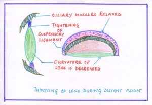
In distant vision, (more than 6 metres) the ciliary muscles are relaxed and the lens becomes flatter or thinner due to stretching of the suspensory ligaments.
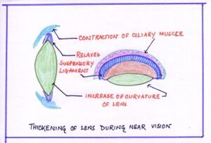
In near vision, (less than 6metres) the ciliary muscles contract pulling the choroid forward, towards the lens and tension is released on the suspensory ligaments. The lens becomes shortened, more convex and thickened due to its elastic nature.
In normal condition the ciliary muscles and are relaxed and the lens remain stretched by the sensory ligament. Then, it is less convex and best suited for the distant vision.
STEREOSCOPIC VISION:- Humans have two eye, both are frontally placed on the face. When both eyes focus on the same object, each eye produces a slightly different image as each eye views from different angles. Stereoscopic vision occurs when for the same object , each eye forms it own separate image of the object on the retina and the vision fields of both the eyes overlap in the center.
Here are some events for the stereoscopic vision:-
- The brain correlates the two images and resolves it into one image only.
- The stereoscopic vision depends on where the eye are located.
- If the eyes are more frontally located , the stereoscopic vision is greater.
- The perception of distance and depth is due to simultaneous focusing of an object in both the eyes and overlapping gives a three-dimensional image.
ADVANTAGES OF STEREOSCOPIC VISION:-
- Stereoscopic vision gives larger visual field than monocular vision( vision by single eye)
- It gives increased accuracy of size and perception of depth and distance of object.
WORKING OF EYE
The working of eye is similar to that of a camera except that the lens of human eye does not move forward or backward but adjusts the distance for focus by undergoing changes in its shape.
PHOTORECEPTION
| EYE | PHOTOGRAPHIC CAMERA |
| Lense muscle | Camera focusing aperture |
| Eyelid | Lense cap |
| Eye ball | Camera box |
| Pupil | Lense aperture |
| Lense | Lense |
| Iris | Diaphragm |
| Retina | Photographic film or plate |
- When the light rays from any object fall on cornea, they enter the eye through pupil.
- Iris controls the amount of light passing through by controlling the size of pupil.
- The rays of light converge as they pass through the cornea, aqueous humour, lens and vitreous humour and finally focus at retina forming an inverted image of the object.
- The light rays are bent at the cornea and the lens makes the final adjustment to bring a sharp focus on retina.
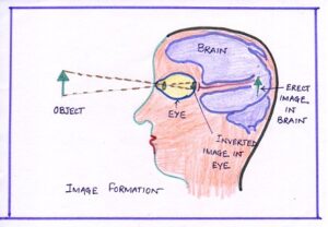
The inverted image formed at the retina is picked by the optic nerve which takes it to the brain where it is reversed and finally an upright image of the object is perceived by the individual.
COMMON DEFECTS OF VISION
| NAME OF THE DEFECTS | SYMPTOMS | REASON | CORRECTIVE MEASURE | RELATED DIAGRAM |
| Myopia or near-sightedness | i)Person cannot see distant objects clearly
ii)The person can see nearby objects clearly. |
i)Eyeball is lengthened
ii)Lens is too much curved or too convex . |
Use of concave lense. | 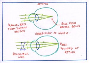 |
| Hypermetropia or long sightedness | Person cannot see nearby objects clearly. | i)Eyeball is shortened.
ii)lens is too flat or less curved. |
Use of biconvex lense | 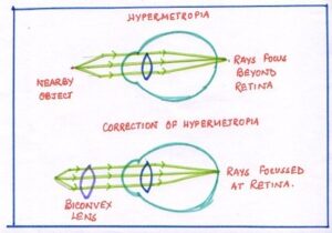 |
| Astigmatism | Formation of blurred image . | Uneven curvature of cornea | Use of cylindrical lense. | 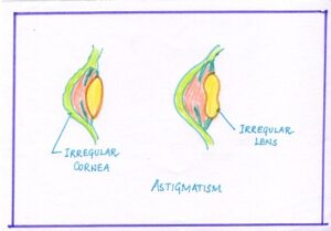 |
| Presbyopia (old age long sightedness) | Affects older people (after 40) cannot see distant or nearby object clearly. | i)Loss of flexibility of lens.
ii)Failure of accommodation. |
Use of bifocal lense. | No Image |
| Cataract or sofia motia | Hazy vision. | Lens becomes opaque. | Surgical removal of lens and implantation of artificial lens or use of spectacles with convex lens. | No Image |
| Glucoma or kala motia | Person cannot see at all. | i)secretion of aqueous humour is increased.
ii)intraocular pressure is increased. |
Cured by drugs if detected on time. | No Image |